Ovarian Hyperstimulation Syndrome (OHSS) is a potentially serious complication that can occur in women undergoing fertility treatments, particularly in the context of assisted reproductive technologies such as in vitro fertilization (IVF). Monitoring and early detection of OHSS are crucial for effective management and prevention of severe complications. Ultrasound imaging plays a pivotal role in assessing the ovarian response and identifying the characteristic features associated with OHSS.
Ultrasound Imaging in Ovarian Hyperstimulation Syndrome:
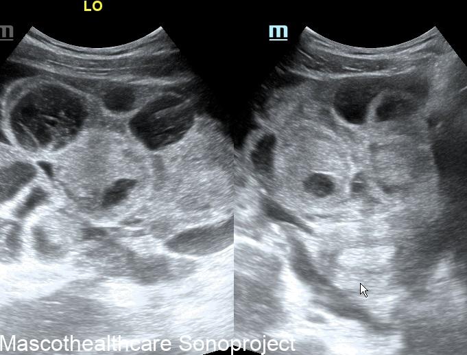
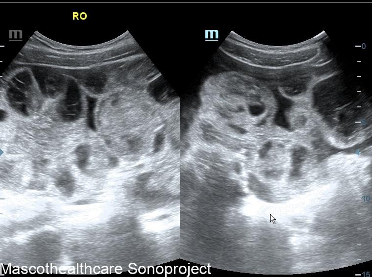
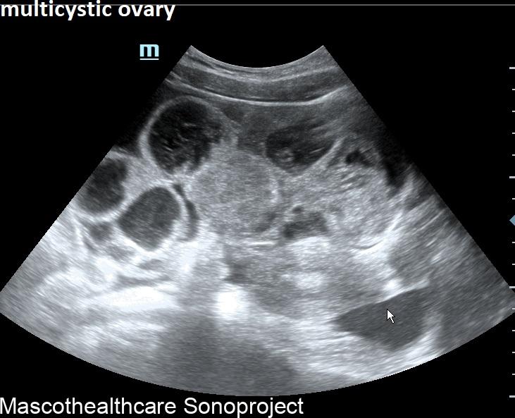
- Ovarian Enlargement: One of the primary ultrasound features of OHSS is the significant enlargement of the ovaries. During controlled ovarian stimulation, multiple follicles develop in response to hormonal medications. In OHSS, these follicles can become excessively enlarged, leading to enlarged ovaries. Ultrasound allows for the measurement of ovarian size, aiding in the assessment of the severity of OHSS.
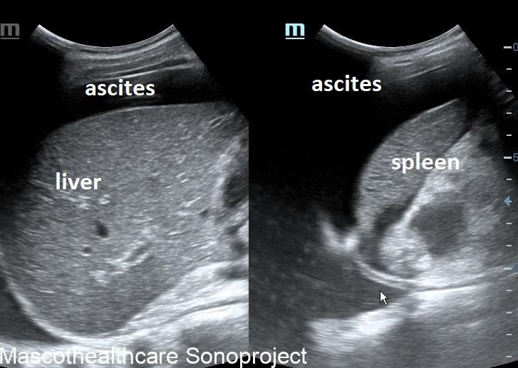
- Ascites: Ascites, the accumulation of fluid in the abdominal cavity, is a common manifestation of moderate to severe OHSS. Ultrasound imaging helps visualize the presence and quantify the amount of ascitic fluid. Ascites can lead to increased abdominal girth and discomfort, and its early detection through ultrasound is crucial for timely intervention.
- Pleural Effusion: In severe cases of OHSS, fluid accumulation may extend beyond the abdominal cavity to involve the pleural space around the lungs. Ultrasound can detect pleural effusion, another important marker of advanced OHSS. Pleural effusion can contribute to respiratory distress, emphasizing the need for careful monitoring through imaging.
- Ovarian Cysts: The development of ovarian cysts is a common consequence of ovarian stimulation. Ultrasound allows for the identification and characterization of these cysts. In OHSS, the cysts may be more numerous and larger than expected, contributing to the overall ovarian enlargement. There may be hemorrhagic changes within the cyst, demonstrated as echo-rich cystic components.
- Clinical Grading: Ultrasound findings are often used in conjunction with clinical symptoms to grade the severity of OHSS. The Golan and Navot criteria, for example, incorporate ultrasound parameters such as ovarian size, ascites, and pleural effusion to categorize OHSS into mild, moderate, and severe forms.