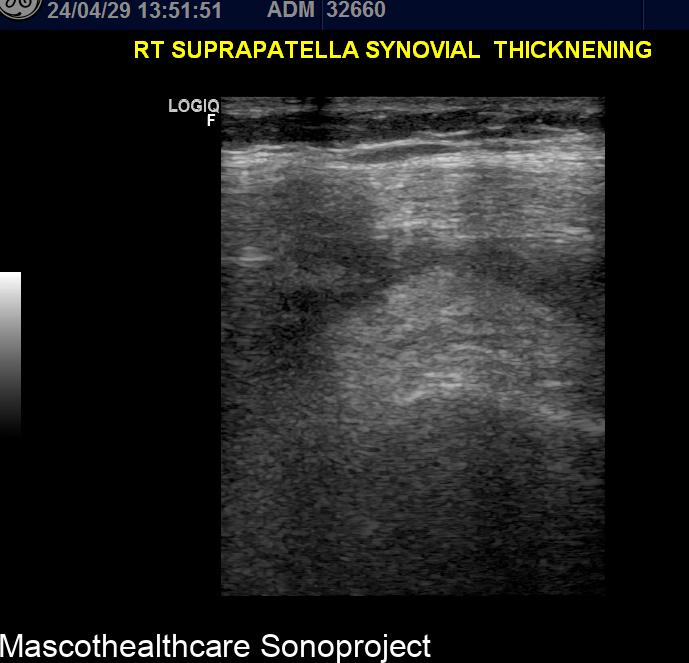
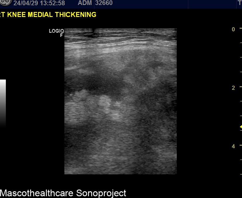
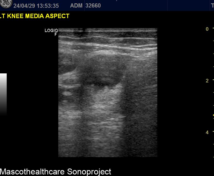
Pigmented villonodular synovitis (PVNS) is a condition characterized by abnormal growth of the synovial tissue in joints. While MRI is often the preferred imaging modality for PVNS, ultrasound can be a useful tool for initial evaluation and monitoring. Here's a look at some key ultrasound features of PVNS:
- Joint effusion: Ultrasound detects moderate effusion within the joint; this may be anechoic or contain debris echoes.
- Synovial thickening: The synovial membrane appears thickened. The thickened synovium may develop finger-like projections (villi) or nodules, which protudes into the effusion spaces.
- Hypoechoic masses: PVNS can cause the formation of nodules within the joint. These masses typically appear hypoechoic and fairly circumscribed.
- Increased vascularity: Doppler ultrasound may reveal increased blood flow within the thickened synovium and masses, suggesting active disease.
It's important to note that these findings are not specific to PVNS and can be seen in other joint conditions such as chronic synovitis, tuberculous synovitis, Giant cell tumor and Rheumatoid arthritis . However, ultrasound can be a quick and relatively inexpensive way to raise suspicion for PVNS, prompting further evaluation with MRI for a more definitive diagnosis.