Duodenal atresia is a congenital condition characterized by the absence or complete closure of a portion of the duodenum, which is the first part of the small intestine. This condition typically presents early in life and is often associated with other congenital anomalies.
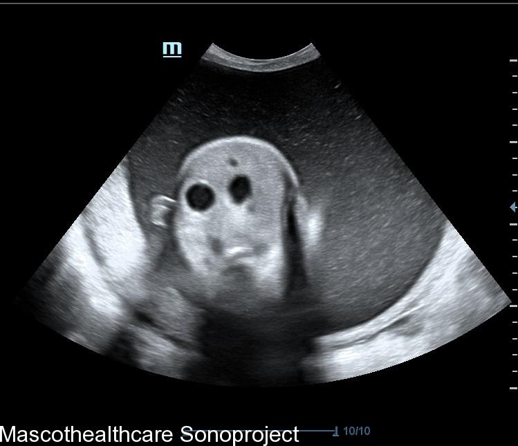
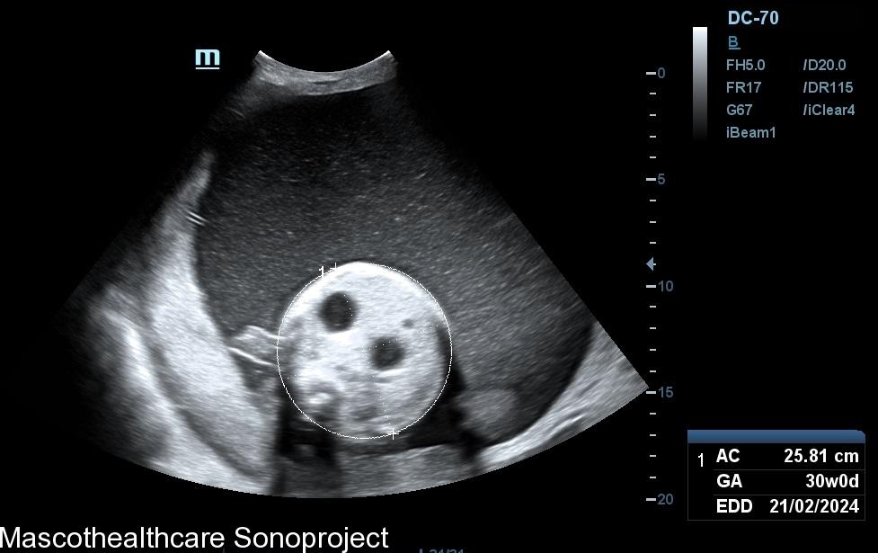
- Double Bubble Sign:
- One of the hallmark ultrasound findings in duodenal atresia is the presence of the "double bubble sign." This refers to the identification of two distinct, dilated fluid-filled structures in the upper abdomen.
- The first bubble represents the stomach, which is often distended due to the accumulation of swallowed amniotic fluid.
- The second bubble corresponds to the dilated proximal portion of the duodenum, which is located just distal to the pylorus.
- Polyhydramnios:
- Duodenal atresia is commonly associated with polyhydramnios, which is an excess accumulation of amniotic fluid. This is a result of the obstruction in the gastrointestinal tract, preventing the normal passage and absorption of amniotic fluid by the fetus.
- Absence of Fluid Distally:
- Beyond the dilated portion of the duodenum, there is often an absence or significant reduction in the amount of amniotic fluid in the distal small bowel.
- This absence of fluid distally is due to the obstruction caused by the atretic segment, preventing the normal flow of amniotic fluid through the duodenum and into the rest of the small intestine.
- Associated Anomalies:
- In addition to evaluating the duodenum, ultrasound may also reveal associated anomalies. Duodenal atresia is frequently seen in conjunction with other congenital conditions such as trisomy 21 (Down syndrome), cardiac anomalies, and renal abnormalities.
- Serial Scans for Confirmation:
- Serial ultrasound scans may be performed to monitor the progression of duodenal atresia and assess for any changes in the appearance of the double bubble sign. This can be especially useful for determining the severity of the obstruction and planning for postnatal management.
It is important to note that while ultrasound is a valuable tool for the prenatal diagnosis of duodenal atresia, additional imaging modalities such as fetal MRI may be employed to provide more detailed information about the anatomy and associated anomalies. The definitive diagnosis and planning for appropriate medical or surgical interventions are often made after birth, with additional imaging and clinical assessments.
Early detection and management are crucial for the optimal care of infants with duodenal atresia.