Liver cirrhosis, the irreversible scarring of liver tissue, presents a significant global health challenge. Early diagnosis is crucial for managing complications and improving patient outcomes. While liver biopsy remains the gold standard, ultrasound imaging offers a safe, non-invasive, and readily available alternative for initial assessment and disease monitoring.
Sonography's Diagnostic Toolbox:
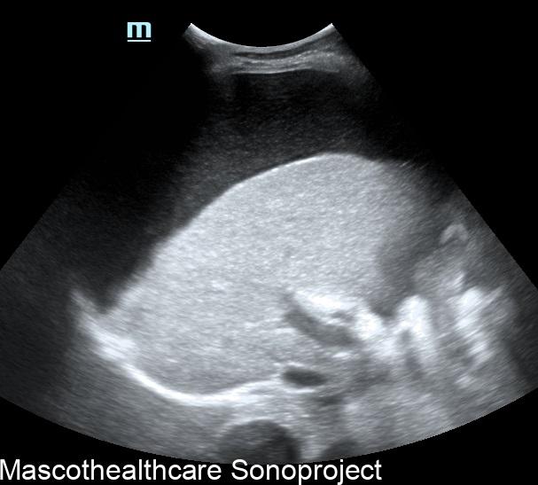
- Liver size and shape: In advanced cirrhosis, the liver shrinks and develops a rounded edge, losing its sharp borders. There may relative caudate lobe sparing.
- Parenchymal texture: Healthy liver tissue appears homogenous, while cirrhotic livers exhibit increased echogenicity (brightness) with a coarse, nodular texture and outline due to scar tissue.
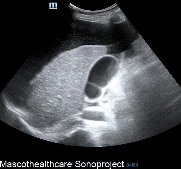
- Vascular changes: Portal vein diameter increases, while hepatic vein flow diminishes, indicating portal hypertension, a hallmark of cirrhosis. Portal vein flow may be reversed (flows backwards towards the feet)in severe cases.
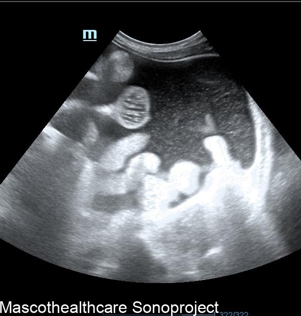
- Spleen enlargement: Splenomegaly, an enlarged spleen, often accompanies cirrhosis due to increased portal pressure.
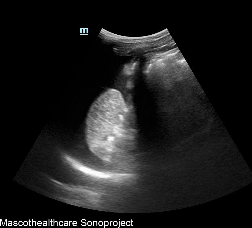
- Identification of nodules: This may be seen in nodular regeneration or malignant transformation
Advanced Techniques for a Sharper Look:
Beyond conventional B-mode imaging, advanced sonographic techniques offer deeper insights:
- Doppler ultrasound: Evaluates blood flow patterns, helping assess portal hypertension and potential complications like variceal bleeding.
- Elastography: Measures tissue stiffness, which increases with fibrosis and scar tissue, aiding in staging cirrhosis severity.
- Contrast-enhanced ultrasound (CEUS): Uses microbubbles to enhance vascularity, improving lesion detection and differentiation between cirrhosis and other liver conditions.
Strengths and Limitations:
Sonography boasts several advantages:
- Safe and painless: No radiation exposure, making it suitable for repeated examinations and monitoring disease progression.
- Widely available: Readily accessible in most healthcare settings.
- Cost-effective: More affordable compared to other imaging modalities like CT or MRI.
However, limitations exist:
- Operator dependence: Image quality and interpretation rely on the sonographer's expertise.
- Limited tissue characterization: Cannot definitively differentiate cirrhosis from other liver diseases with similar sonographic features.
- Challenges in obese patients: Excess body fat can impede sound wave penetration, hindering image quality.
The Bigger Picture:
Sonographic imaging plays a vital role in the initial assessment and management of liver cirrhosis. While not a replacement for liver biopsy in all cases, it provides valuable information for diagnosis, disease staging, and monitoring treatment response. As technology advances, with improved resolution and tissue characterization capabilities, sonography's role in diagnosing and managing liver cirrhosis is poised to become even more significant.