Ultrasound imaging, often associated with imaging of the abdomen and pelvis, has a growing role in chest imaging, offering a non-invasive and radiation-free modality for evaluating various thoracic conditions. While ultrasound is not the primary imaging choice for certain structures within the chest due to limitations in penetrating air-filled structures like the lungs, it has valuable applications in specific scenarios.
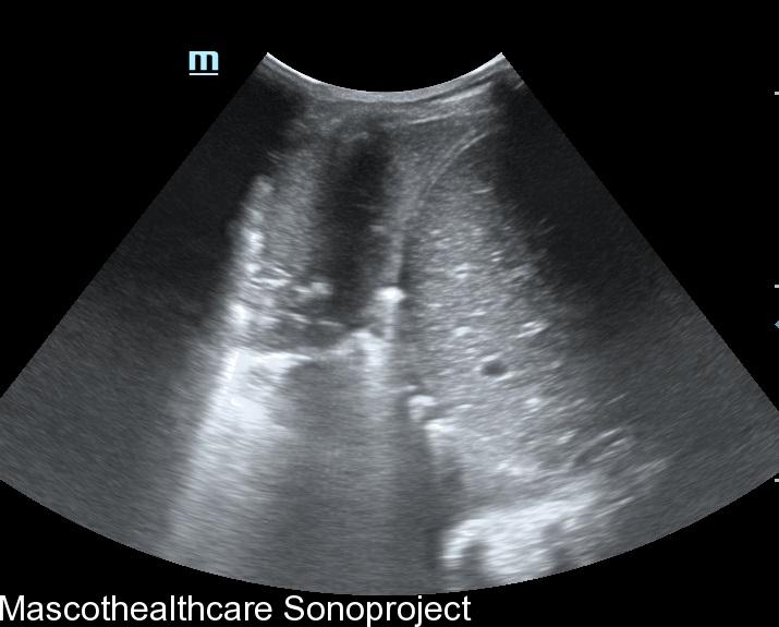
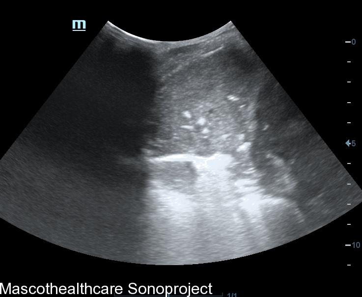
- Pleural and Pericardial Effusions:
- Ultrasound is highly effective in detecting and characterizing pleural and pericardial effusions. It can visualize fluid collections in the pleural and pericardial spaces, helping to guide drainage procedures such as thoracentesis or pericardiocentesis.
- Pneumothorax Assessment:
- Ultrasound plays a crucial role in the rapid assessment of pneumothorax, especially in emergency situations. It can identify the presence of air in the pleural space and help distinguish between simple pneumothorax and more severe conditions.
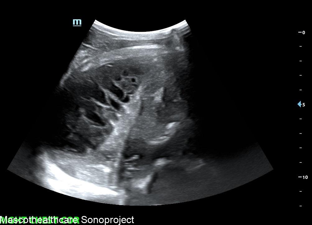
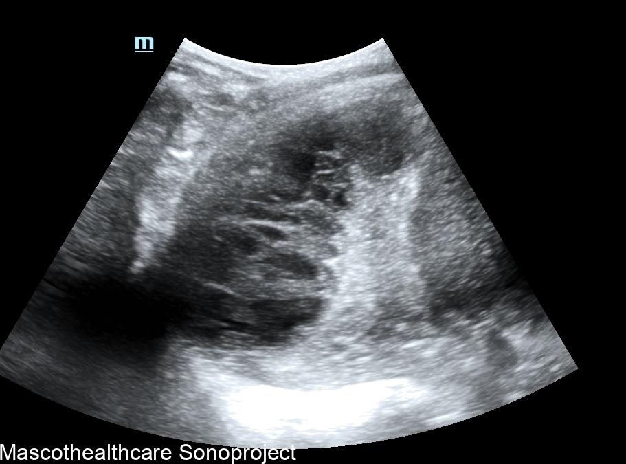
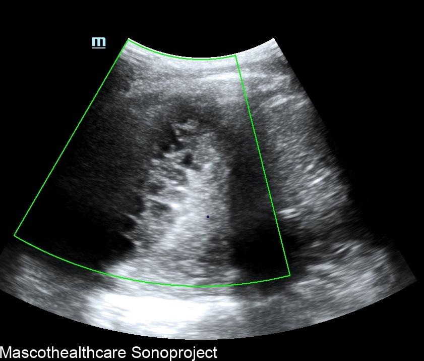
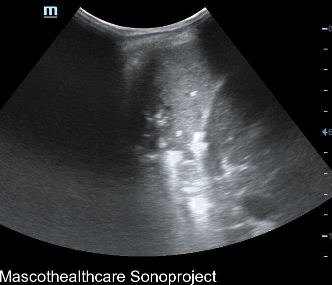
- Evaluation of Lung Consolidations:
- While air-filled structures like the lungs limit the direct visualization of lung parenchyma, ultrasound can be used to evaluate peripheral lung consolidations. It is particularly useful in identifying subpleural lesions or assessing the pleura adjacent to consolidated lung tissue, especially in children.
- Guidance for Procedures:
- Ultrasound guidance is commonly employed for various chest procedures, including thoracentesis, paracentesis, and lung biopsies. Real-time visualization of the needle insertion site helps enhance accuracy while minimizing the risk of complications.
- Thyroid and Neck Structures:
- The upper part of the chest often includes structures such as the thyroid and neck vessels. Ultrasound is a valuable tool for evaluating thyroid nodules, lymph nodes, and vascular structures in the neck, providing detailed information without exposure to ionizing radiation.
- Pediatric Chest Imaging:
- Ultrasound is particularly useful in pediatric chest imaging due to its lack of ionizing radiation. It can aid in assessing congenital abnormalities, chest masses, lung consolidation or pleural effusions in children.
While ultrasound has its strengths in specific applications, it is important to acknowledge its limitations, particularly in imaging structures that contain air, such as the lungs. Complementary imaging modalities like chest X-rays and CT scans remain essential for comprehensive evaluation of thoracic conditions. The role of ultrasound in chest imaging continues to evolve, and ongoing technological advancements may expand its applications further in the future.