Sonographic evaluation of the appendix is commonly performed to make evaluation of the appendix, rule out appendicitis and other causes of acute abdomen. It is particularly useful in children and pregnancy where the exposure to radiation is eliminated.
- Patient Preparation: Typical patient present with abdominal pain, nausea, vomiting or fever. Such cases won't require any preparation.
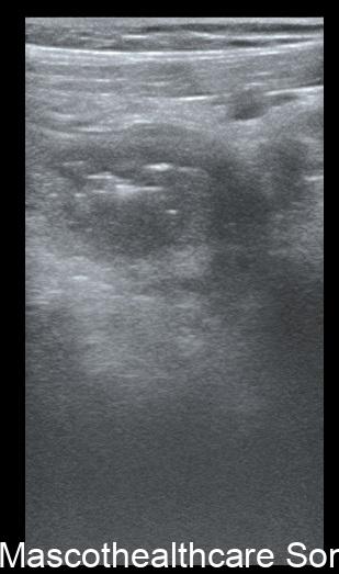
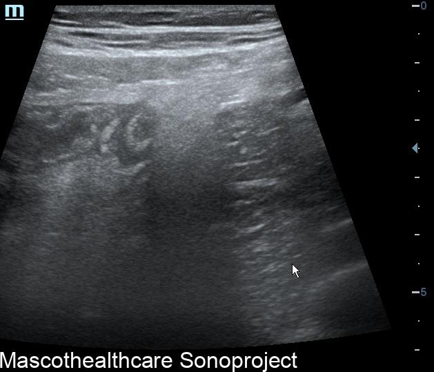
- Transducer and Technique: A high-frequency ultrasound transducer is used for the examination. Lower frequency probes are also useful for individuals with thicker anterior abdominal wall adipose. The transducer is placed on the right lower quadrant while using graded compression to optimize the visualization, which is the area where the appendix is normally located(except in rare cases of Situs or malrotation). Visibility of the appendix is better with inflammation as normal appendix are more difficult to visualize.
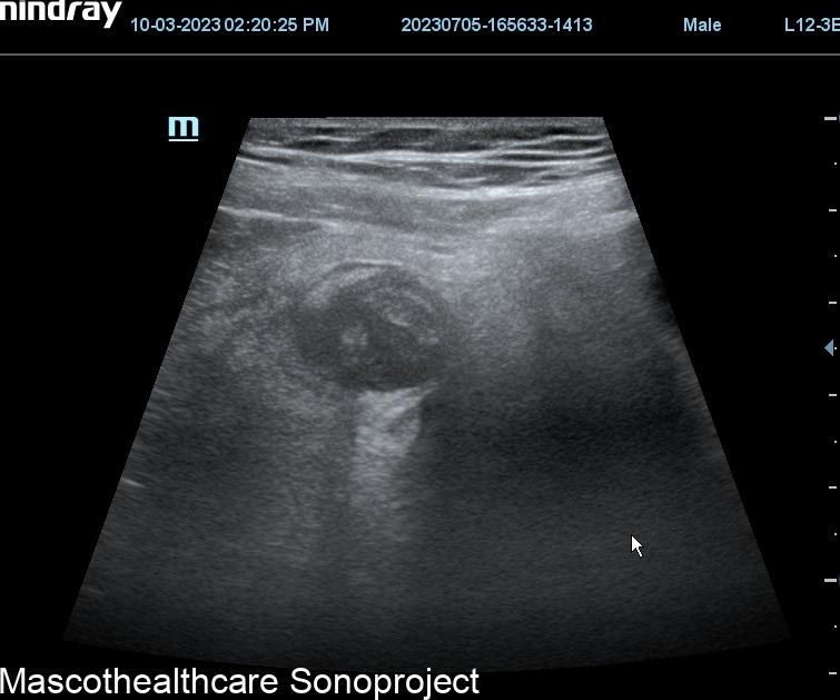
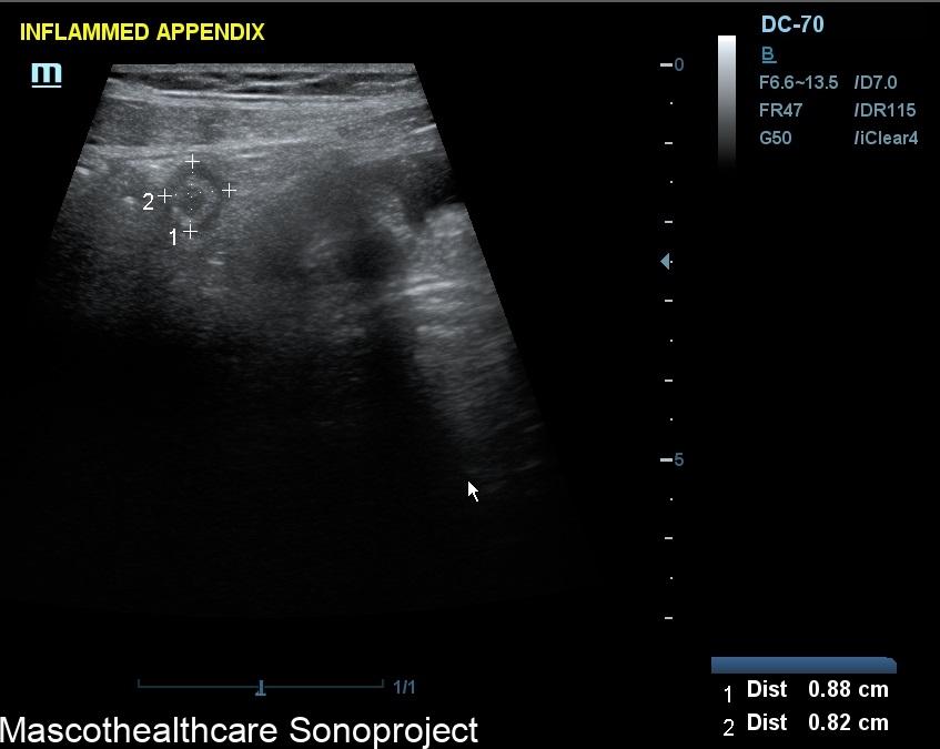
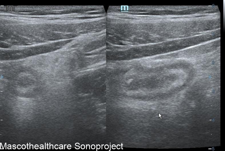
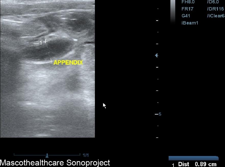
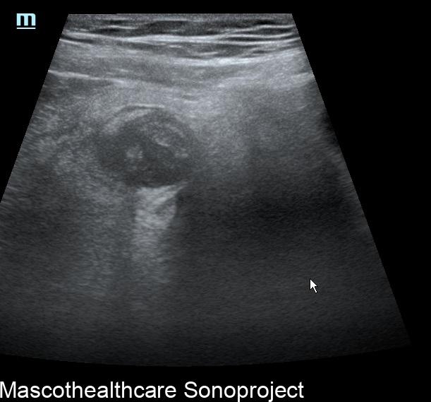
- Identification of the Appendix: The normal appendix is a blind-ended, tubular structure arising from the cecum. It is typically located in the right lower quadrant of the abdomen and may extend to the pelvis or retrocecum. The cecum is identified and the appendix is traced from its base to its tip. The appendix is often visualized as a compressible, tubular structure with a diameter of less than 6 mm.
- Sonographic Signs of Appendicitis: Sonographic signs of appendicitis may include:
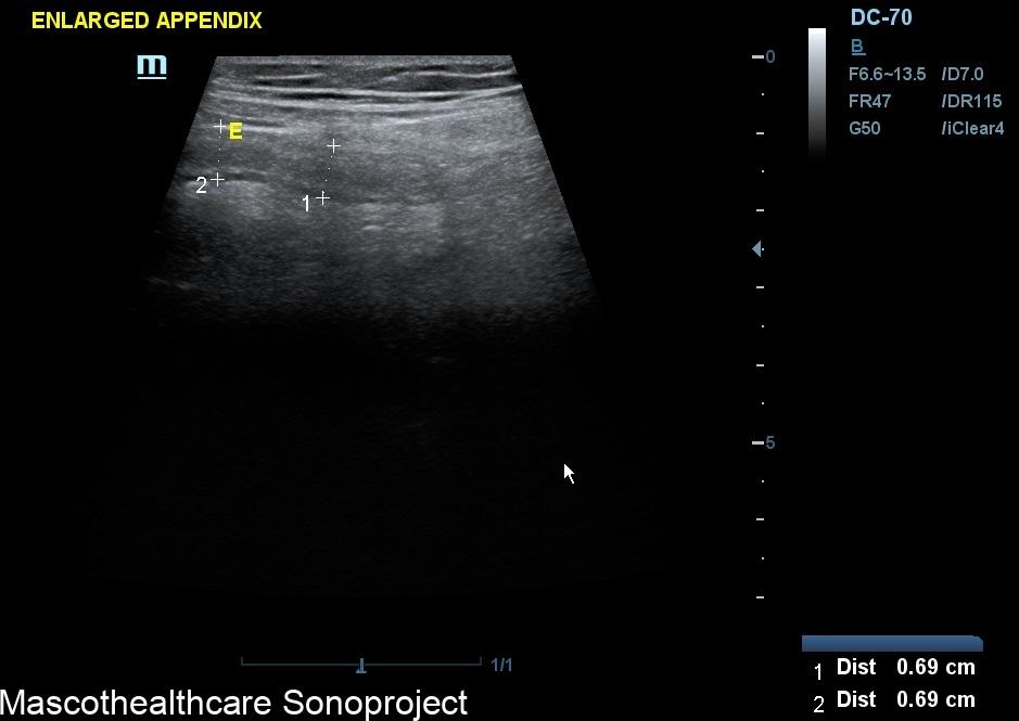
- Increased diameter of the appendix (greater than 6 mm is suggestive).
- Marked probe tenderness on the appendix.
- Non-compressibility of the appendix.
- Presence of echogenic (bright) surrounding fat.
- Peri-appendiceal fluid collection.
- Hyperemia of the appendix on color Doppler imaging.
- Secondary Signs: In addition to direct signs of appendicitis, one may also look for secondary signs such as increased blood flow around the inflamed appendix or the presence of an appendicolith (a calcified deposit within the appendix).
- Complications: Deeply hypoechoic appendix or patchy loss of the appendiceal gut signature are typical findings in gangrenous appendicitis. Other features are peri-appendiceal fluid collection and discontinuity of appendiceal wall indicating appendiceal perforation. Collection of echo-rich fluid with or without gaseous pockets are seen in appendiceal abscess. Irregular heterogenous mass.with thickened adjacent bowel wall and echogenic mesentery suggests a phlegmon. The identification of these complications is crucial for appropriate clinical management.
- Limitations: It's important to note that while ultrasound is a valuable tool, it does have limitations. Operator-dependency of ultrasound and the need for a skilled sonographer in obtaining accurate results.In some cases, factors such as patient body habitus or overlying bowel gas may limit visualization of the appendix. In these situations and in cases of high clinical suspicion and negative findings, additional imaging modalities like computed tomography (CT) may be considered.