Ultrasound technology has come a long way since its inception, offering a wide range of applications in the field of medicine and beyond. While many people associate ultrasound with pregnancy scans, there are various types of ultrasounds, each tailored to specific purposes. In this comprehensive guide, we will explore the different types of ultrasounds and their diverse uses in healthcare and other industries.
1. 2D Ultrasound
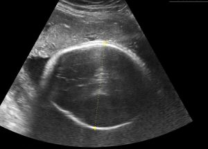
Principle: Two-dimensional ultrasound, often referred to as 2D ultrasound, is the most basic and commonly used type of ultrasound. It uses a single transducer to emit sound waves, which bounce back to create a flat, black-and-white image of the internal structures.
Applications: 2D ultrasounds are used for various purposes, including monitoring fetal development during pregnancy, diagnosing abdominal and pelvic conditions, and evaluating blood flow.
2. 3D Ultrasound
Principle: Three-dimensional ultrasound, or 3D ultrasound, uses multiple transducers to capture a series of 2D images from different angles. A computer then assembles these images into a three-dimensional representation.
Applications: 3D ultrasounds provide more detailed and realistic images of the fetus during pregnancy, aiding in the diagnosis of congenital abnormalities. They are also used in gynecology, urology, and cardiology for better visualization of organs and structures.
3. 4D Ultrasound
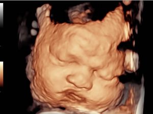
Principle: Four-dimensional ultrasound, also known as 4D ultrasound, is an extension of 3D ultrasound that adds the dimension of time. It creates moving, real-time 3D images, allowing for dynamic visualization of the fetus or internal organs.
Applications: 4D ultrasounds are primarily used during pregnancy to provide parents with a more vivid and realistic view of their developing baby's movements and expressions. They can also aid in diagnosing fetal abnormalities.
4. Doppler Ultrasound
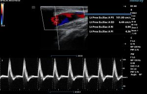
Principle: Doppler ultrasound uses the Doppler effect to assess blood flow in arteries and veins. It measures the frequency shift of sound waves bouncing off moving blood cells to create color-coded images or waveforms.
Applications: Doppler ultrasounds are essential in vascular studies to diagnose conditions like deep vein thrombosis, carotid artery disease, and peripheral artery disease. They are also used in obstetrics to monitor blood flow in the umbilical cord and fetal vessels.
5. Transesophageal Echocardiogram (TEE)
Principle: TEE is a specialized type of ultrasound used to assess the heart. A transducer is inserted into the esophagus to obtain high-resolution images of the heart and its structures.
Applications: TEE is used to evaluate heart valve function, congenital heart defects, blood clots, and cardiac anatomy. It is particularly useful during cardiac surgeries and interventions.
6. Endoscopic Ultrasound (EUS)
Principle: Endoscopic ultrasound combines ultrasound with endoscopy. A small ultrasound probe is attached to an endoscope and inserted into the body through natural openings or small incisions.
Applications: EUS is primarily used in gastroenterology for visualizing the gastrointestinal tract, pancreas, and nearby structures. It aids in the diagnosis and staging of digestive system disorders and cancers.
7. Musculoskeletal Ultrasound
Principle: Musculoskeletal ultrasound focuses on imaging muscles, tendons, ligaments, and joints. It is often used in sports medicine and orthopedics.
Applications: Musculoskeletal ultrasound helps diagnose conditions like tendonitis, ligament injuries, and joint inflammation. It is also used for guided injections and aspirations in the musculoskeletal system.
Conclusion
Ultrasound technology has evolved significantly, offering a range of imaging options for various medical specialties and applications. Each type of ultrasound serves a unique purpose, providing healthcare professionals with valuable insights and patients with a means to monitor and diagnose medical conditions. As technology continues to advance, the capabilities of ultrasound imaging continue to expand, leading to improved healthcare outcomes and a deeper understanding of the human body.